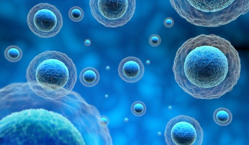
Bioanalytical assays such as ELISA cytokine assays have been used successfully to assess preclinical and clinical evaluations such as pharmacokinetics studies. In addition to ELISA assay development, custom ELISA assay development has further helped researchers tailor their experimental approach. Along with bioanalytical assays, cell-based assay development has been widely conducted in drug discovery and development. Historically, two-dimensional (2D) cell cultures were the only viable cell-based screening options. 2D cell models can effectively predict drug responses in vivo. However, they have several disadvantages associated with biochemical and mechanical cues, loss of tissue-specific architecture, and cell-to-matrix and cell-to-cell interactions.
However, the advent of 3D cell cultures has accelerated the productivity of drug research and development. Using 3D cell cultures combined with cell models such as primary cells and stem cells allows greater predictability of toxicity and safety before the drug moves to the clinical trial phase. This mode of operation further reduces the attrition rate of novel medicines under development. The current article explores the incorporation of 3D cell culture models into cell-based assay development and how it may improve the predictive power of drug discovery.
3D cell culture models improving the predictive power of drug discovery studies
Recent advances in tissue engineering, microfabrication approaches, and cell biology have helped develop multiple 3D cell culture technologies. These technologies include organoids, multicellular spheroids, scaffolds, hydrogels, 3D bioprinting, and organs-on-chips. Each of these techniques has its advantages and disadvantages. Although these 3D technologies are different in protocol and principle, they all restore morphological, microenvironmental, and functional characteristics of human tissues and organs. Moreover, drug discovery is a lengthy and complex process. 3D cell cultures have been used in all aspects of the drug discovery process, including disease modeling, target identification, validation, screening, efficacy, and safety profiles.
Generally, drug discovery begins with a clinical condition or a disease without a suitable medication. Today disease modeling has become increasingly vital for fulfilling unmet therapeutic needs. As 3D cell cultures can bridge the gap between 2D cultures and in vivo analysis, they are employed to evaluate the mechanisms of different diseases. 3D cell cultures have gained prominence, particularly in tumor biology, as traditional 2D cultures lack in addressing issues of metastatic colonization, indolent disease, relapse, dormancy, and evolution of drug resistance.
During preclinical drug discovery, target identification and validation often slow down the entire process. However, 3D cultures have the potential to identify new mechanisms and targets and also accelerate the identification and validation of targets. The primary reason for these advantages is that gene expressions in 3D cell culture models are closer to in vivo models compared to 2D monolayer cultures.
Cell-based screening is usually the start to identify hit compounds in early drug discovery. As HTS-compatible cellular systems are simple, low-cost, and highly efficient, target-based HTS has dominated the hit identification process. However, recent years have witnessed the growth of phenotypic screening. Although combining 3D cultures with HTS systems is still in its infancy, they show potential in identifying clinically relevant compounds and enabling effective preclinical and clinical research.
Must Read: The Role of ELISA Services in Drug Discovery and Development: A Critical Component of the Pipeline
Multiplexed ELISA for Biomarker Discovery: Uncovering New Insights in Cancer Research